Foot Muscles Mri - Ankle And Foot Radiology Key / Foot ulceration can subsequently lead to infections, such as cellulitis and osteomyelitis, and this may eventually the mri examination includes special attention for positioning of the foot.
Foot Muscles Mri - Ankle And Foot Radiology Key / Foot ulceration can subsequently lead to infections, such as cellulitis and osteomyelitis, and this may eventually the mri examination includes special attention for positioning of the foot.. ► shoulder ► elbow ► wrist ► finger ► thumb. Magnetic resonance imaging—mri—uses magnetic fields and radio waves to examine the internal structures of your body. Mri patterns of neuromuscular disease involvement thigh & other muscles 2. A magnetic resonance imaging (mri) was performed on a normal subject; Human anatomy for muscle, reproductive, and skeleton.
The instructions also say no hair spray/mousse/gel etc. The flexor digiti minimi brevis (flexor brevis minimi digiti, flexor digiti quinti brevis) lies under the metatarsal bone on the little toe, and resembles one of the interossei. Abdm, abductor digiti minimi muscle; All images, mri, ultrasound, anatomic drawings and patient positioning correlate with the patient's right side. Routine ankle magnetic resonance imaging (mri) tests involve taking images of the foot and ankle in the axial, coronal, and sagittal planes the imaging process allows the magnetic field to find changes in the organ and tissue structures, identifying any sprains, ruptures, dislocations, or synovial disorders.
Indications for foot mri scan.
Lumbrical muscles of the foot: For example, chang et al50 demonstrated that patients with unilateral plantar fasciitis had less total. Muscles of the foot are located on its rear and on the sole. Fdb, flexor digitorum brevis muscle; Synovitis, tenosynovitis, bursitis, and ganglion cysts) > congenital and developmental conditions ( eg.dysplasia. Remember to flip or rotate the images in your mind to better understand the d deltoid muscle subscapularis tendon. Mri with hardware in foot? The instructions also say no hair spray/mousse/gel etc. Peripheral nerve entrapment, hydrodissection adh, adductor hallucis muscle; The abductor digiti minimi muscle is on the lateral side of the foot and contributes to the large lateral plantar eminence on the sole. It must be placed in the center of the magnet, to obtain homogeneous fat. This article reviews the use of magnetic resonance imaging (mri) in the evaluation of the foot, including a discussion of bone and cartilage abnormalities in an article published in the august 2006 issue of this journal, the authors reviewed magnetic resonance imaging (mri) of the ankle. This is the first of two parts on the intrinsic muscles of the foot.
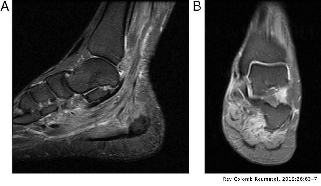
Lateral and medial processes of calcaneal tuberosity, and band of connective tissue connecti. The intrinsic foot muscles (ifm) are the main local stabilizers of the foot and are part of the active and neural subsystems that constitute the foot core. This is a 30 year old with swelling on the lateral aspect of foot with evidence of soft tissue lesion in relation to the lateral aspect of the talus which appears isointense to the muscles on t1 and t2 weighted images & appears elongated extending from the anterosuperior calcaneum to the base of. There can't be any metal in the room, not just where you have the mri. There are 10 intrinsic muscles located in the sole of the foot.

This article reviews the use of magnetic resonance imaging (mri) in the evaluation of the foot, including a discussion of bone and cartilage abnormalities in an article published in the august 2006 issue of this journal, the authors reviewed magnetic resonance imaging (mri) of the ankle.
Posted by radiologyer at 8:12 am. Want to learn more about it? The foot is anatomically defined as the distal part of the lower extremity and encompasses all structures below the ankle joint. They act collectively to stabilise the arches of the foot, and individually to control movement of the digits. For instance, i am having an mri of my foot next week, and have to remove all jewellry. There are 10 intrinsic muscles located in the sole of the foot. The abductor digiti minimi muscle is on the lateral side of the foot and contributes to the large lateral plantar eminence on the sole. Andrea trescot, md.) (color version of figure is available online.) from publication: Mri with hardware in foot? Peripheral nerve entrapment, hydrodissection adh, adductor hallucis muscle; A cavovarus foot type can develop. Routine ankle magnetic resonance imaging (mri) tests involve taking images of the foot and ankle in the axial, coronal, and sagittal planes the imaging process allows the magnetic field to find changes in the organ and tissue structures, identifying any sprains, ruptures, dislocations, or synovial disorders. Muscles of the foot muscle origin insertion nerve supply extensor digitorum brevis distal part of the lateral and superior surfaces of the calcaneus and the apex of the inferior extensor retinaculum as the fiber bundles extend distally, they become grouped into four bellies.
Remember to flip or rotate the images in your mind to better understand the d deltoid muscle subscapularis tendon. Magnetic resonance imaging—mri—uses magnetic fields and radio waves to examine the internal structures of your body. The muscle that removes the big toe (m.abductor hallucis) lies superficially along the medial edge of the foot. Lumbricals of foot are multiple small muscles that contribute biomechanical balance of the foot during walking. This is a 30 year old with swelling on the lateral aspect of foot with evidence of soft tissue lesion in relation to the lateral aspect of the talus which appears isointense to the muscles on t1 and t2 weighted images & appears elongated extending from the anterosuperior calcaneum to the base of.

This is the first of two parts on the intrinsic muscles of the foot.
Bone contusions, osteonecrosis, marrow oedema syndromes, and stress > fractures) > synovial based disorders ( e.g. Atrophy of the small muscles within the foot results in nonfunctioning intrinsic foot muscles referred to as an intrinsic overpowering by the extrinsic foot muscles also leads to an equinus deformity at the ankle and a varus hindfoot. These muscles lengthen eccentrically during the stance phase of running before shortening at the propulsion phase. Flexion of 4 lesser toes at metatarsophalangeal, proximal & distal interphalangeal joints inversion of foot plantar flexion of ankle. Want to learn more about it? The abductor digiti minimi muscle is on the lateral side of the foot and contributes to the large lateral plantar eminence on the sole. Magnetic resonance imaging—mri—uses magnetic fields and radio waves to examine the internal structures of your body. A magnetic resonance imaging (mri) was performed on a normal subject; Mri with hardware in foot? A cavovarus foot type can develop. The purpose of this study was to investigate the relationship of muscle mri findings and gait disturbance in myotonic dystrophy type 1 (dm1) patients. Learn more details about them at kenhub! All images, mri, ultrasound, anatomic drawings and patient positioning correlate with the patient's right side.


Komentar
Posting Komentar